
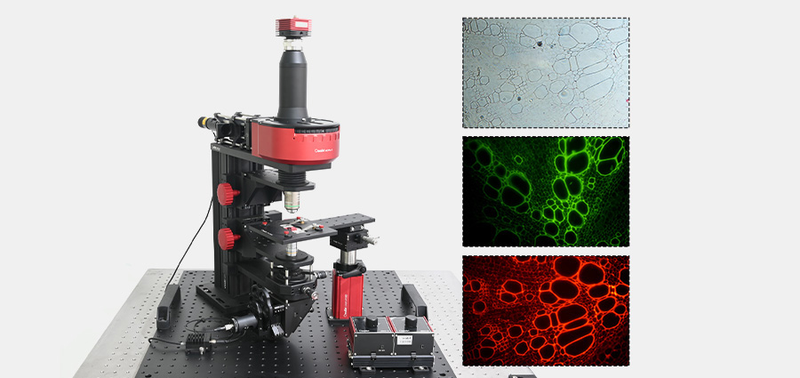
OMBA-EFM Epi-Fluorescence Microscope
Technical description: What is fluorescence microscopy? Experiment overview: Fluorescence microscopes examine organic and inorganic samples using fluorescence and phosphorescence to reveal structural organization and spatial distribution, particularly in complex specimens unsuitable for conventional transmitted-light microscopy. Two types are available. Transmitted fluorescence microscopy resembles ordinary microscopy: light travels from the base through the condenser to excite the sample, then through the objective to the eyepiece. The more common epi-fluorescence microscope directs excitation through the objective onto the specimen and collects emitted fluorescence back through the objective to the eyepiece. Procedure: (1) Excitation: A high-intensity xenon lamp, mercury lamp or laser produces selected wavelengths directed through filters onto the specimen. (2) Emission: Fluorescent dyes or proteins absorb excitation energy, enter an excited state and return to the ground state by emitting longer-wavelength fluorescence. (3) Filtering/imaging: An emission filter removes residual excitation and passes selected fluorescence to an eyepiece or camera to form a clear image. Objectives: Based on substances emitting a different wavelength after absorbing light, fluorescence microscopy is widely used in biological research to study intracellular uptake/transport and chemical distribution/localization. (1) Specific molecular labeling/observation: Fluorescent probes reveal proteins, DNA, RNA and other structures and their distribution/dynamics. (2) High-contrast imaging: Unlabeled regions normally do not emit, reducing background and providing high SNR and target contrast. (3) Live-cell dynamics: Real-time observation of cell division, transport and signaling in living cells or tissues. (4) Multiplex/multicolor imaging: Different dyes simultaneously label multiple targets, showing relative positions and interactions. (5) Three-dimensional imaging: Combined with techniques such as confocal microscopy, scanning different focal planes builds 3D images. Kit list: Contact customer service for the detailed contents. Experimental content.
 Save to Favorites
Overview
Key Features
Datasheet
Please provide your email to download the datasheet.
Invalid email format. Please try again.
Supplier Information
Related Products
-
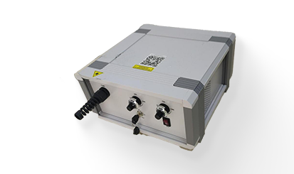 LWMS-T3
Microscopes
Laser-wave
Wavelength: Any 3 wavelengths from 450,488,520,635,660,785 Wavelength Deviation: ±5 nm Power: 10-100 mW
-
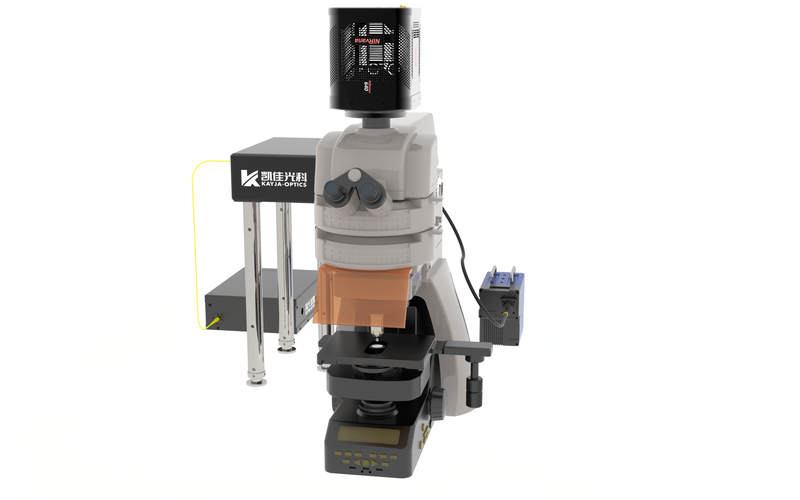 Second Near-Infrared Window SIM Imaging System
Microscopes
Kayjaopt
NIR-II multifocal structured-illumination super-resolution microscopy system. By integrating multifocal structured illumination microscopy (MSIM) into fluorescence microscopy in the NIR-II region (1000-1700nm), the system achieves NIR-II MSIM super-resolution imaging for the first time. In a highly scattering medium, it reaches spatial resolution of nearly 1.5um at a depth of 2.5mm. Further results at longer emission wavelengths show excellent performance. NIR-II MSIM is a powerful imaging method combining deep penetration with enhanced spatial resolution and contrast.
-
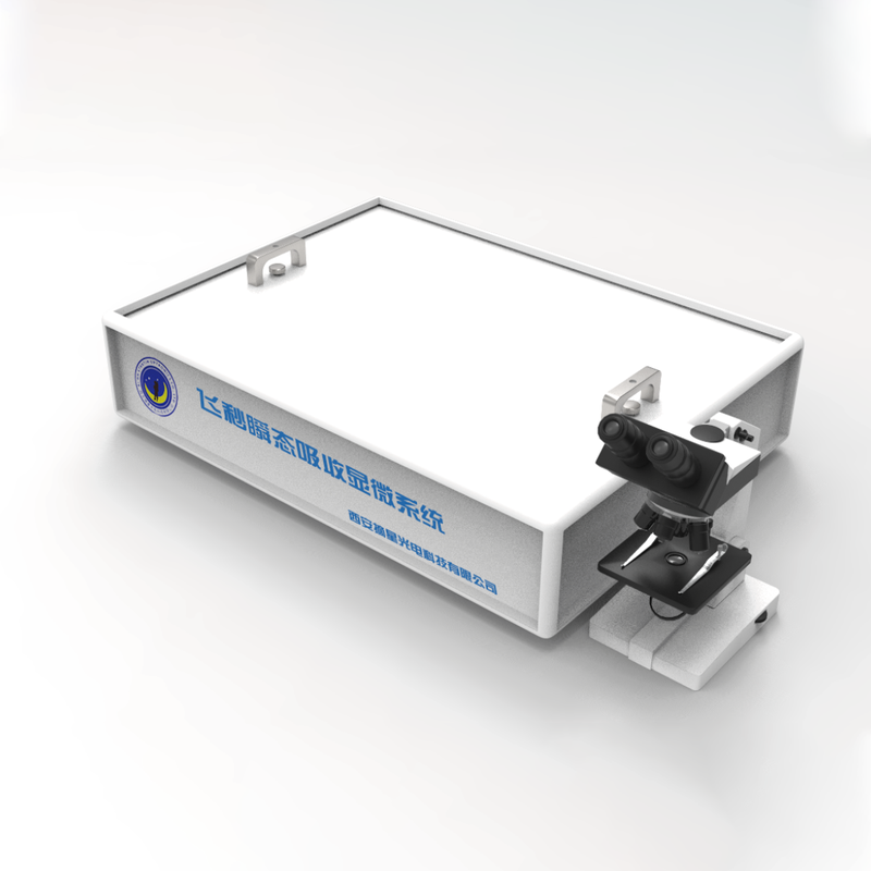 Femtosecond transient absorption microscopy system
Microscopes
Startin-optronics
This system combines an Olympus inverted microscope and a high-speed CMOS camera to visualize photoexcited-carrier dynamics in micro/nano samples. It also measures microscopic transient reflection/transmission spectra and dynamics, with femtosecond time resolution and spatial resolution <1μm. The complete system includes pump-probe optics, microscopy, spectral and image acquisition, a data-processing computer and software. It can work with cryogenic, magnetic-field, high-pressure and TCSPC equipment.
-
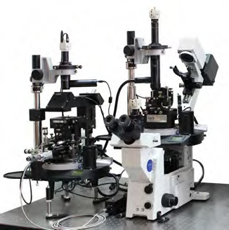 Scanning probe microscope
Microscopes
Startin-optronics
Scanning Probe Microscopy (SPM) encompasses probe techniques developed from scanning tunneling microscopy, including atomic-force, electrostatic-force, magnetic-force, scanning-ion-conductance and scanning-electrochemical microscopy. These surface-analysis instruments integrate optoelectronics, lasers, weak-signal detection, precision mechanics, automatic control, digital signal processing, applied optics, high-speed computer acquisition/control and high-resolution graphics. Applications span physics, chemistry, biology, medicine, materials and microelectronics in research and industry.
Related Articles
As a core precision optical component for wavelength screening, frequency locking and spectral demodulation, optical etalons are widely used in high-end scientific research and precision laser equipment.
As a vital emerging sector of modern three-dimensional spatial economy, the low-altitude economy has become a new global growth driver. It covers a wide range of commercial scenarios, including UAV navigation and obstacle avoidance, precise airspace management, aerial mapping, low-altitude logistics distribution, security monitoring and emergency disaster rescue. Nevertheless, frequent extreme weather, complex terrain obstacles and increasingly dense low-altitude flight activities pose severe challenges to the safety and stability of low-altitude aircraft operations.
Laser pump sources are the core energy input component of solid-state lasers and laser amplifiers, and their performance directly determines the output power, beam quality and service life of the entire laser system. In scenarios such as industrial processing, scientific research experiments and high-end equipment, improper pump source selection will not only result in substandard system performance, but also bring a series of problems such as rapid device degradation and high operation and maintenance costs. This article sorts out the core selection logic and key pitfalls of pump source procurement, and provides selection references based on mature, mass-produced high-reliability products to help purchasers efficiently screen suitable products.
Shandong Haucore Optoelectronics Co., Ltd. introduces the HGLD‑792‑COS‑D‑10WB semiconductor laser within its COS series. Delivering an output power of 10 W at a central wavelength of 792 nm, this device adopts gold‑tin bonding COS packaging. Featuring high reliability and long service life, it serves as a preferred pump source for solid‑state lasers and is widely deployed in industrial processing, laser display, medical & aesthetic treatment, scientific research and other fields.
Loading...
Name
Phone
Organization
Use Case

Database Platform