
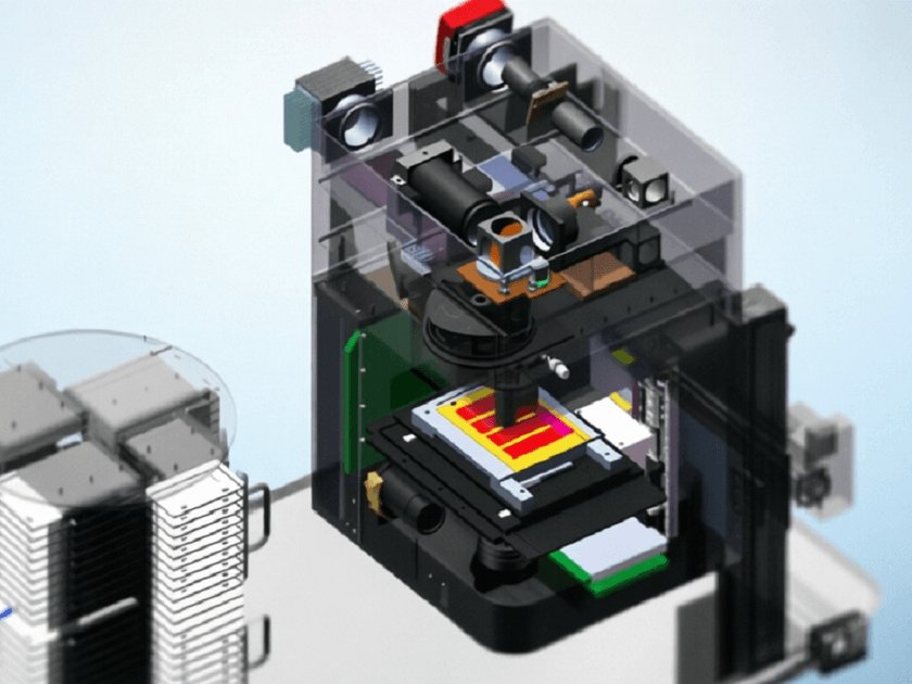
Hyperspectral Pathology Slide Analysis Microscope
Hyperspectral Pathology Slide Analysis Microscope Hyperspectral imaging technology upgrades traditional microscopes from 'morphological observation' to 'composition analysis'. Our hyperspectral pathology microscope integrates a liquid crystal tunable filter (LCTF) or fiber spectrometer into the bright-field/fluorescence microscopy optical path, achieving pixel-by-pixel spectral acquisition of pathological slides (covering 400nm-1000nm with spectral resolution better than 2nm). By analyzing the characteristic spectra of different tissue types (such as cancer cells and normal cells), it assists doctors in label-free, automated pathological grading and boundary recognition. The system beam path uses Kohler illumination and infinity-corrected design to ensure full-field illumination uniformity and spectral consistency; the automatic scanning stage supports full-slide hyperspectral imaging, combined with deep learning algorithms to generate pathological auxiliary diagnosis maps. This equipment can significantly improve the diagnostic efficiency of pathological slides of the digestive tract, breast, lung and other tissues and reduce subjective interpretation differences. It is suitable for pathology departments of Class A tertiary hospitals, third-party medical laboratories and pharmaceutical companies' drug histology evaluation platforms.
 Save to Favorites
Overview
Applications
Key Features
Datasheet
Please provide your email to download the datasheet.
Invalid email format. Please try again.
Supplier Information
Related Products
-
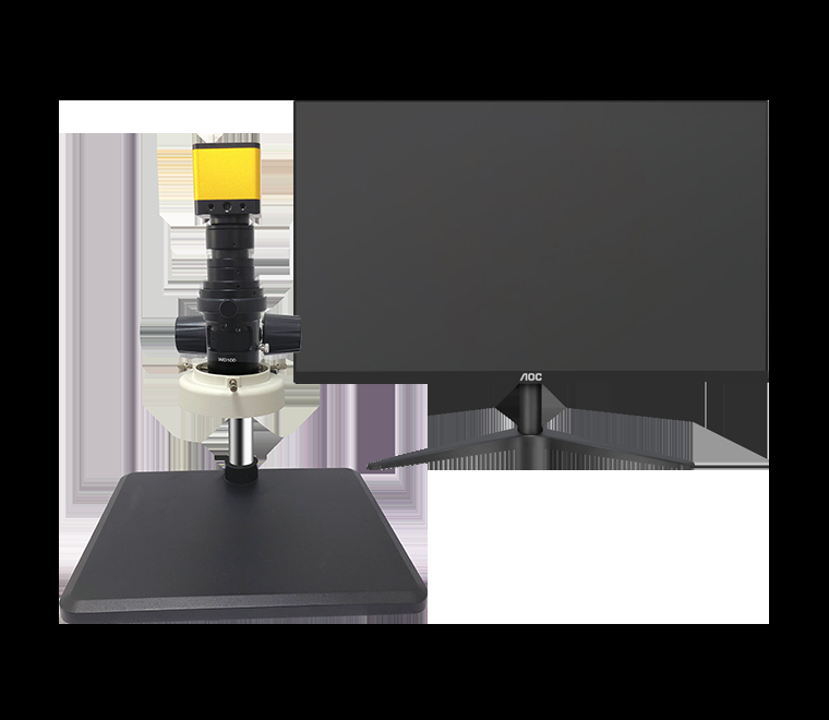 CT-200 microscope
Microscopes
Sunsightimage
CT-200 microscope
Microscopes
Sunsightimage
Optical Dimensions: 1/3 Optical magnification multiplier: 100X Objective Lens: 1X
-
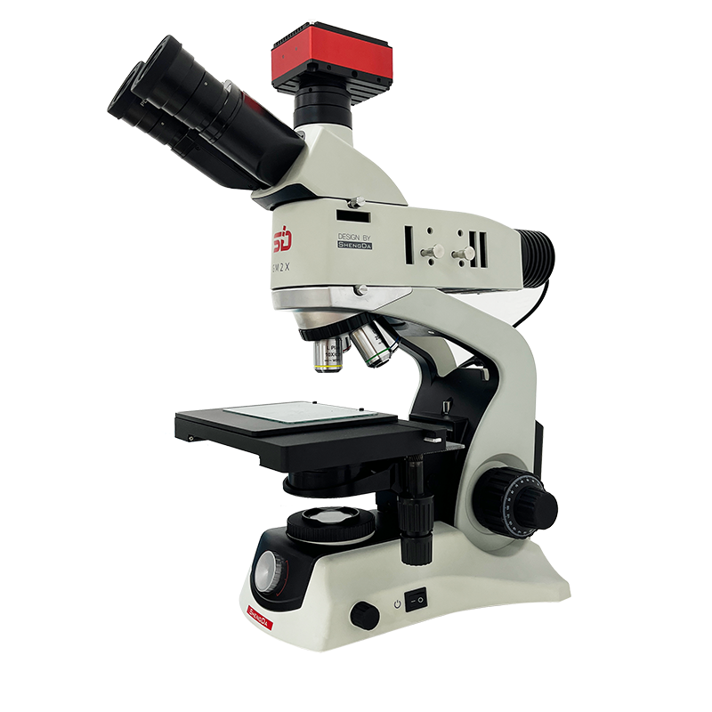 GM2X gold microscope
Microscopes
Sunsightimage
GM2X gold microscope
Microscopes
Sunsightimage
-
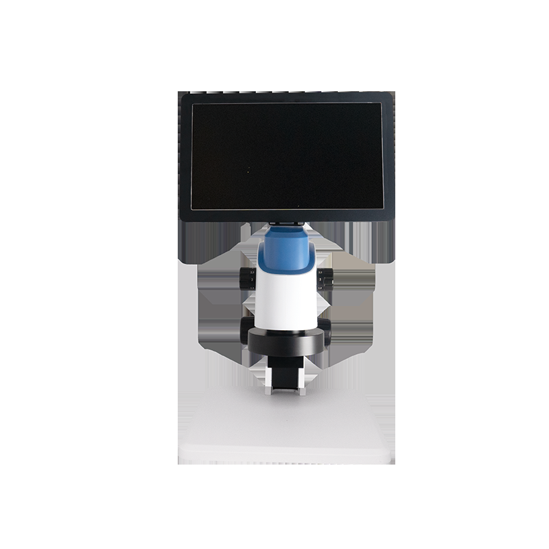 HT-8068 microscope
Microscopes
Sunsightimage
HT-8068 microscope
Microscopes
Sunsightimage
Optical Dimensions: 1/2
Dolphin HT8068 video microscope;
-
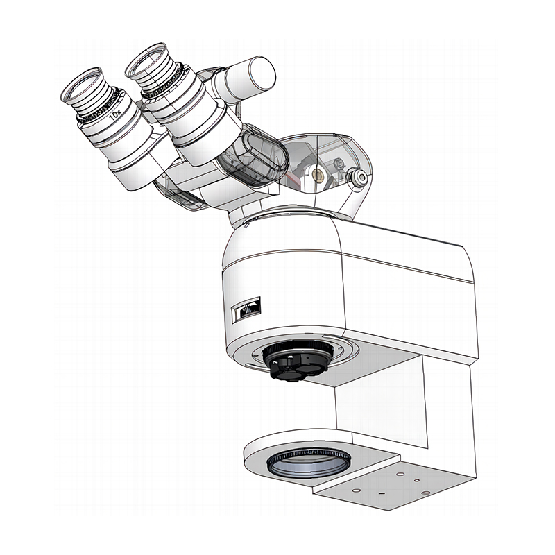 Ophthalmic Operating Microscope – 0.4-2.5X Zoom, 9-52mm Field of View
Microscopes
Risingeo
Ophthalmic Operating Microscope – 0.4-2.5X Zoom, 9-52mm Field of View
Microscopes
Risingeo
Independently developed by RISING EO;
-
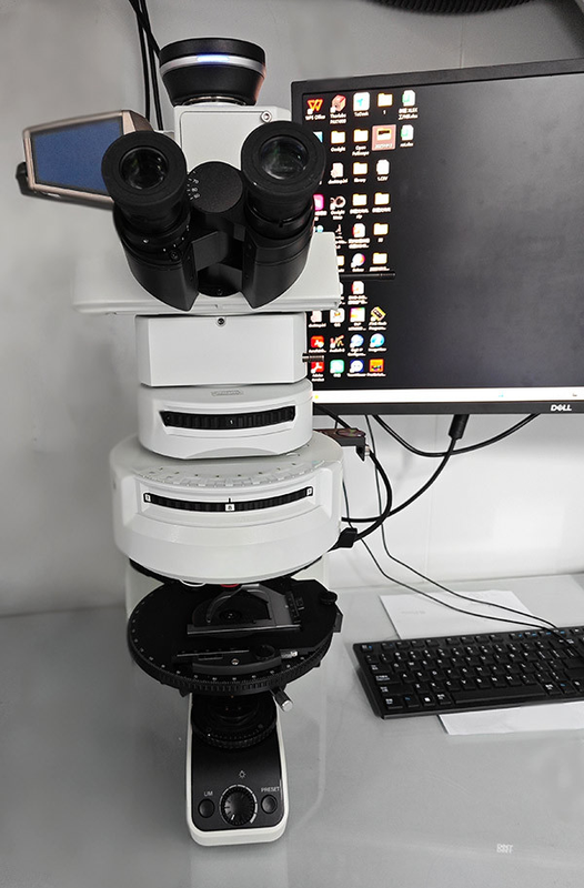 JC-Bire double-reflection microscope
Microscopes
Jc-ultrafast
JC-Bire double-reflection microscope
Microscopes
Jc-ultrafast
JC-Bire double-reflective microscopes; optical microscopes mounted on self-study imaging systems; based on oscillation microtechnologies; compensation optical systems through unique liquid crystals; qualitative and quantitative imaging of double-reflective structures at 546 nm wavelengths
Related Articles
Polycom Photonics Co., Ltd focuses on the R&D and mass production of high-stability, narrow-linewidth laser sources for optical sensing applications. The company provides cost-effective and high-precision optical sensing lasers with a wavelength coverage from 689nm to 2350nm. Featuring accurate wavelength control and outstanding spectral stability, these laser products are widely applied in atomic spectrum detection, element precision measurement and professional optoelectronic sensing systems.
Laser pump sources are the core energy input component of solid-state lasers and laser amplifiers, and their performance directly determines the output power, beam quality and service life of the entire laser system. In scenarios such as industrial processing, scientific research experiments and high-end equipment, improper pump source selection will not only result in substandard system performance, but also bring a series of problems such as rapid device degradation and high operation and maintenance costs. This article sorts out the core selection logic and key pitfalls of pump source procurement, and provides selection references based on mature, mass-produced high-reliability products to help purchasers efficiently screen suitable products.
A spatial modulator, most commonly known as a Spatial Light Modulator (SLM), is a core programmable optical device widely used in modern photonics, imaging, and laser systems. It enables precise, dynamic control of light beams, making it indispensable for advanced optical technology and industrial optical equipment.
Founded in 2003, BWT Beijing Optoelectronics adheres to the mission "Let Dreams Fly With Light" and vision of becoming a Global Leader in Laser Solutions, guided by the core value "Innovate to Achieve Extraordinary".
Loading...
Name
Phone
Organization
Use Case

Database Platform