
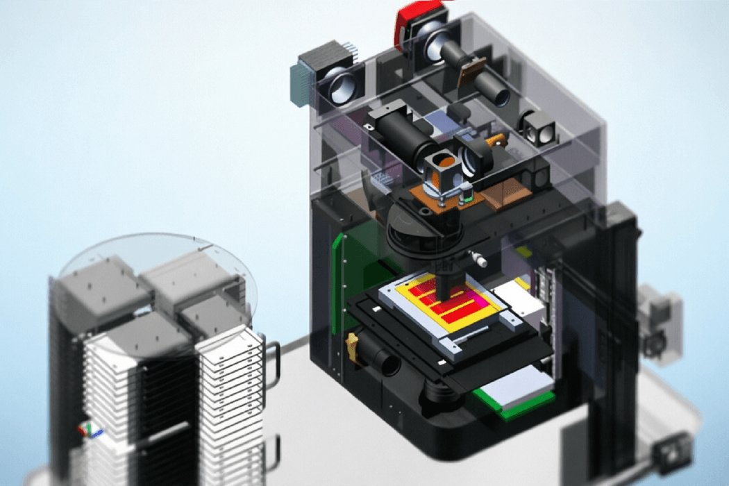
Hyperspectral Pathology Analysis Microscope Objective
Hyperspectral Pathology Analysis Microscope Objective Hyperspectral microscopic imaging upgrades traditional pathological morphology observation to dual 'morphology + chemical composition' analysis. We provide partners with dedicated objectives and optical path solutions suitable for hyperspectral imaging systems — using infinity-corrected achromatic design with a magnification chromatic aberration of ≤0.3% across the wide spectral range of 400nm-1000nm, ensuring precise alignment of images in different bands. The objective NA reaches up to 0.75 with a working distance of 1mm, and can be used with liquid crystal tunable filters or imaging spectrometers. By acquiring the characteristic spectrum of each pixel on pathological slides, normal tissue and cancerous areas can be distinguished label-free, and the distribution of specific proteins or metabolites can be identified. Our objectives have been applied in automatic pathological grading research of tissue sections of the digestive tract, breast, lung and other organs, significantly improving diagnostic efficiency and consistency. We also support integration with automatic scanning stages and spectral library comparison algorithms, helping partners build hyperspectral pathology analysis systems with independent intellectual property and advancing digital pathology toward the molecular level.
 Save to Favorites
Overview
Applications
Key Features
Datasheet
Please provide your email to download the datasheet.
Invalid email format. Please try again.
Supplier Information
Related Products
-
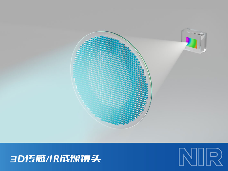 3D /IR Optical Lens
High-Performance Lenses
Metalenx
3D /IR Optical Lens
High-Performance Lenses
Metalenx
As the receiving end of the MetaToFTM series;
-
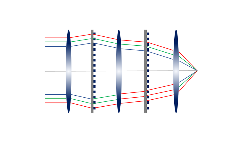 Hybridmeta® mobile-phone lens
High-Performance Lenses
Metalenx
Hybridmeta® mobile-phone lens
High-Performance Lenses
Metalenx
Based on phototracies transfer techniques;
-
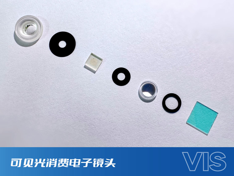 Visible-Light Consumer Electronics Lens
High-Performance Lenses
Metalenx
Visible-Light Consumer Electronics Lens
High-Performance Lenses
Metalenx
Visible photoclipse hybrid systems in consumer electronics;
-
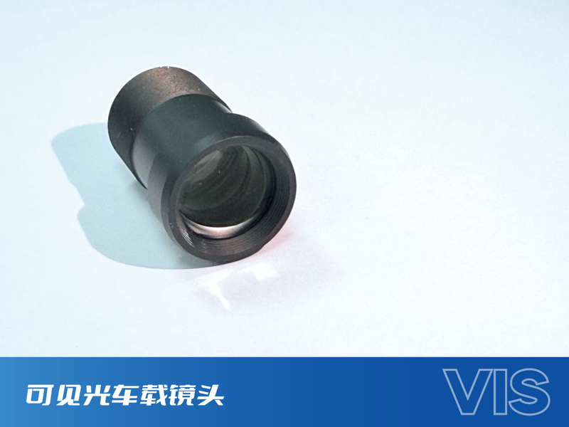 Visible-Light Automotive Lens
High-Performance Lenses
Metalenx
Visible-Light Automotive Lens
High-Performance Lenses
Metalenx
The release by Metallans of the global first wide-spectrum ADAS-8M pre-vision visual optic lenses;
-
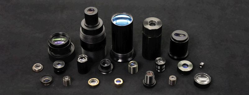 Optical Lens
High-Performance Lenses
Alphaoptics
Optical Lens
High-Performance Lenses
Alphaoptics
Optical lenses are an essential part of the formation of optical systems.
Related Articles
As a professional manufacturer of precision optical thin-film components, sycco-optics (Shandong Yanggu Hengjing Optoelectronics) launches high-performance 850nm narrow band pass filters for near-infrared precision detection, intelligent sensing and optical imaging systems. Adopting advanced vacuum coating technology, the filter realizes high transmittance in the target band and deep blocking in interference bands. It effectively eliminates stray light and spectral noise, serving as a core optical component for machine vision, biochemical detection and photoelectric sensing equipment.
Wuhan Optics Valley Quantum Technology Co., Ltd. is a leading domestic developer of high‑speed semiconductor optical chips and photodetector components. Our product portfolio covers InGaAs avalanche photodiode bare dies and PIN photodetector chips designed for fiber‑optic communication
A microlens array (MLA) is a high-precision optical component composed of hundreds or thousands of tiny, uniformly arranged micro lenses on a single flat substrate. Unlike traditional single lenses that focus one light beam, a microlens array can split, homogenize, and reshape light on a micro scale. It is widely paired with VCSELs, lasers, and image sensors, becoming a core part of modern miniature optical systems.
Shandong Huaguang Optoelectronics Co., Ltd. (HAUCORE) is a leading high-tech manufacturer of compound semiconductor optoelectronic devices, founded in 1999 and headquartered in Jinan, China, with two R&D and production bases in Jinan and Weifang.
Loading...
Name
Phone
Organization
Use Case

Database Platform